
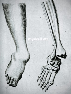
Pott’s fracture, sometimes called a bimalleolar ankle fracture, is a particular kind of bone damage that affects the ankle joint. The fracture of the tibia’s medial malleolus (the bony protrusion on the inside of the ankle) and the fibula’s lateral malleolus (the bony prominence on the outside of the ankle) is known as Pott’s fracture.
An forceful and frequently severe injury to the ankle joint breaks the bones in this form of fracture. Both malleoli bones are typically disrupted as a result of a violent twist, rotational force, or direct contact on the ankle.
Pott’s fracture characteristics include:
Broken medial malleolus: The tibia forms the bony projection known as the medial malleolus, which is located on the inside of the ankle. This portion of the ankle bone is frequently fractured in Pott’s fracture.
Broken Lateral Malleolus: The fibula forms the lateral malleolus, which is situated on the outside of the ankle. In Pott’s fracture, a fracture in this area coexists with a medial malleolus fracture.
Relevant Anatomy:
Joints:
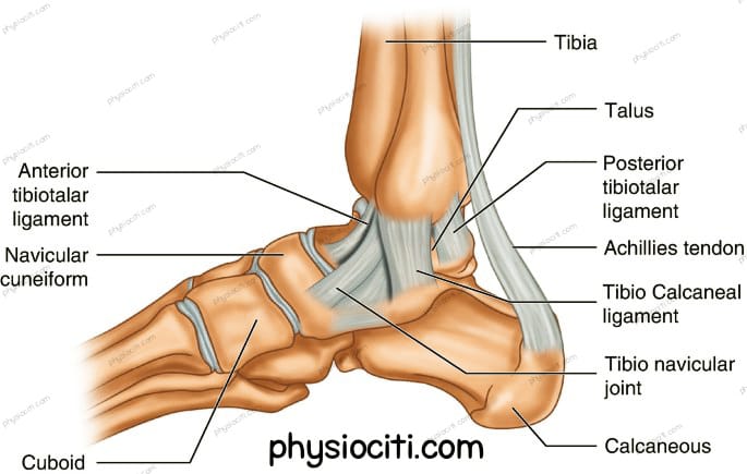
The ankle has three main joints that work together to help with movement and stability:
- Talocrural Joint (Ankle Joint):
- Type: Hinge joint.
- Parts Involved: It’s involves the lower part of the tibia (shinbone) and the upper part of the talus (a bone in the foot).
- Role: Allows up-and-down movement of the foot.
- Inferior Tibiofibular Joint:
- Type: It’s the joint between the ends of the fibula and tibia (the two lower leg bones).
- Supported By: Held together by ligaments called tibiofibular ligaments or syndesmosis.
- Role: Provides stability to the ankle.
- Subtalar Joint:
- Parts Involved: Situated between the talus and the calcaneus (heel bone).
- Divisions: Divided into front and back parts separated by the sinus tarsi.
- Role: Helps in side-to-side movement of the foot.
Ligaments:
The main ligaments connected to the bone prominences implicated in Pott’s fracture are as follows:
1. Medial (Deltoid) Ligaments:
- Description: Located on the inner side of the ankle.
- Components: This ligament complex includes multiple bands: tibionavicular, tibiocalcaneal, posterior tibiotalar, and anterior tibiotalar ligaments.
- Function: Provides stability and prevents excessive outward (eversion) movement of the foot, supporting the inner part of the ankle.
2. Lateral Ligaments:
- Components: The lateral ligament complex consists of: a. Anterior Talofibular Ligament (ATFL): From the front of the fibula to the front of the talus. b. Calcaneofibular Ligament (CFL): Connecting the fibula to the calcaneus (heel bone). c. Posterior Talofibular Ligament (PTFL): Extending from the fibula to the back of the talus.
- Function: These ligaments are important for preventing excessive inward (inversion) movement of the foot. They are commonly injured during ankle sprains, often associated with Pott’s fractures.
3. Syndesmotic Ligaments:
- Description: These ligaments connect the tibia and fibula and support the inferior tibiofibular joint.
- Components: Include the anterior-inferior tibiofibular ligament (AITFL), posterior-inferior tibiofibular ligament (PITFL), and interosseous ligament.
- Function: Maintains stability between the tibia and fibula, restricting excessive movement between these bones.
Although the ankle joint’s bony prominences are the main target of damage in Pott’s fracture, the force or severity of the fracture can also injure the ligaments that surround these bones. Ligament injuries may happen simultaneously with a fracture, impairing the ankle joint’s stability and functionality. Treating ligament injuries is a common part of rehabilitation in order to stabilize and function properly of the ankle.
Mechanism Of Injury of Pott’s Fracture
Pott’s fractures are usually caused by a violent, frequently traumatic event that damages the ankle joint and breaks the bony prominences surrounding the ankle. An outline of the common methods is provided below:
- Rotational or twisting force: The ankle joint’s bones may break if the ankle rotates or twists outside of its typical range of motion.
- Direct Impact: Pott’s fractures can occur from direct hits to the ankle joint, such as falls.
- Fall: Ankle twisting unnaturally might result from falling from a height or slipping on an uneven surface, placing a great deal of stress on the bones and perhaps breaking them.
- Sports Injuries: Sports and physical activities that include rapid movements, jumping, high impact, or direction changes might put people at risk for ankle injuries, such as Pott’s fracture.
- Bone Health and Age: As people age, their bones may deteriorate and become more prone to fractures from less powerful blows or strains.
- Motor vehicle Accidents: Pott’s fracture can happen in certain accident situations, particularly in auto accidents where there is a lot of force or stress to the lower leg.
- Bone Weakening: Weakened by diseases like osteoporosis, bones are more prone to fractures, even those brought on by very small trauma.
“In addition to other injuries such a twisted ankle, ankle dislocation, or other fractures in the foot, ankle, or lower leg, Pott’s fracture frequently occurs. Pott’s fractures can vary widely in kind and intensity. They can include both sides of the ankle (bi-malleolar), be undisplaced (bones remain in place), displaced (bones don’t line up), or even break the skin (compound).
Differentiating between a serious sprain and a fracture is difficult to do visually. Excessive ankle twisting might cause both. They both result in severe pain, edema, and difficulty moving.”
Read More: Sprained ankle vs broken ankle.
Clinical Presentations of Pott’s Fracture
Signs
- Pain: severe and sudden discomfort in the area of the ankle joint. When pressure is put to the injured ankle or when movement occurs, the pain is worse.
- Bruising and Swelling: Due to blood and tissue damage, there is rapid bruising and swelling around the ankle area. Ankle is sensitive to the touch and seems swollen.
- Deformity or displacement: In extreme circumstances, there might be an obvious deformity or misalignment of the ankle joint. Bones could seem misaligned or moved.
- Restricted range of motion: soreness and swelling in the ankle joint causing difficulty in movement. Trying to move the ankle could make it more uncomfortable.
- Difficulty of Bearing Weight: pain and instability in the affected leg, making it difficult or impossible to bear weight on it.
Symptoms
- Difficulty walking: Ankle instability makes it difficult to stand or walk normally. Sensations of instability or incapacity to bear all of one’s weight on the injured leg.
- Tenderness: There may be painful spots to the touch around the ankle, particularly over the bony prominences.
- Weakness and Instability: Weakness and instability in the ankles, which makes it challenging to sustain body weight and keep one’s balance.
- Functional limitations: diminished capacity to carry out regular tasks requiring use of the injured ankle.
Common Disability in Pott’s Fracture
The foot may occasionally twist in a Pott’s fracture, causing it to bend in an unusual manner known as a valgus position. The muscles that support the foot’s arch may be stretched as a result. This type of walking can give the impression that the foot is flatter (FLAT FOOT) than it is when someone has Pott’s fracture. This occurs as a result of stretched-out muscles that are unable to perform their normal function of maintaining the arch of the foot.
Diagnosis:
Medical history & Physical examination
HISTORY: A thorough patient history is necessary for the diagnosis of Pott’s fracture, and an increase in leg diameter at the malleoli level indicates a fibular fracture (Keen’s sign of Pott’s fracture).
PHYSICAL EXAMINATION: The doctor will conduct a thorough physical examination of the ankle, checking for signs of swelling, bruising, tenderness, deformity, and assessing the range of motion and stability of the joint.
Imaging Tests
- X-rays: With an x-ray, the diagnosis of a Pott’s fracture can be verified. Following a diagnosis, it is common practice to categorize the fracture using either the Danis-Weber classification system, which considers the location of the distal fibular fracture in relation to the ankle joint’s syndesmosis, or the Lauge-Hansen classification system, which is based on the rotational mechanism of injury.
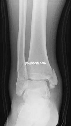
- CT-Scan: When X-rays are unclear or additional evaluation of the fracture pattern is required, a computed tomography (CT) scan may be requested to get more precise images of the fracture.
- MRI (Magnetic Resonance Imaging): An MRI may be suggested to assess ligament or tendon problems related to the fracture in some complicated instances or if soft tissue injury is suspected.
Differential diagnosis
To rule out further complications, the medical professional will take into account other possible injuries or disorders, that may present with identical symptoms.
- Ankle Sprain
- Avulsion Fracture
- Syndesmotic Injury (High Ankle Sprain)
- Ligamentous Injury
- Tendon Injury
- Osteochondral Lesions
- Contusion or Soft Tissue Injury
- Infection or Septic Arthritis
Physiotherapy Management of Pott’s Fracture
Physiotherapy is an essential component of the comprehensive management of Pott’s fracture, with the goal of helping the injured ankle regain its normal strength, function, and mobility. The exact injury severity, healing stage, and functional goals of each individual are essential while designing the physiotherapy program. An overview of physiotherapy management for Pott’s fracture is provided below:
1. Immobilization Phase
Protecting Against Swelling: The injured ankle needs protection, and swelling needs to be controlled initially. Inflammation can be decreased with the use of methods including ice therapy, elevation, and mild movements of the leg’s uninjured parts.
2. Early Mobilization
- RANGE OF MOTION EXERCISES
Ankle alphabet exercise: Using your big toe, trace the alphabet’s letters while maintaining a stable ankle.
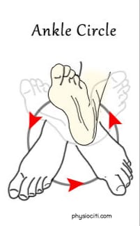
Ankle Circles: To increase flexibility and decrease stiffness, rotate your ankle in both clockwise and counterclockwise directions.
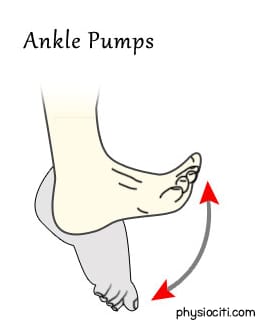
- NON-WEIGHT BEARING ACTIVITIES
Exercises like ankle pumps, toe curls, and light stretching that don’t directly strain the ankle.
3. Strengthening Exercises
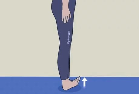
Toe Raises: While keeping your heels firmly planted, slowly lift your toes off the ground while sitting or standing. Lower them back down after holding them for a little while. Repetition should be increased gradually as tolerated.
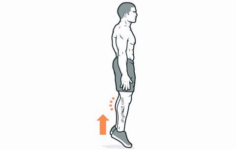
Calf Raises: While standing on both feet, raise your heels off the ground by rising onto your toes. Lower youself back down slowly. As you restore strength, move on to single-leg calf lifts from the starting position of both feet.
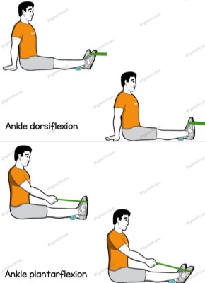
Resistance Band Exercises: Pull your toes toward your shin with an ankle dorsiflexion exercise, or point your toes away from your shin with a plantar flexion exercise, using resistance bands.

4. Balance & Proprioception Exercise
Single-Leg Stance: Balance while standing on the unaffected leg. If necessary, use a support or cling to a sturdy surface. For an extra challenge, try doing this exercise while closed-eye.

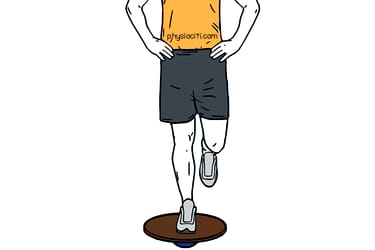
Exercises with a balancing board or wobble board: These devices can be used, under the supervision of a trained specialist, to enhance ankle stability and proprioception.
5. Gait Training
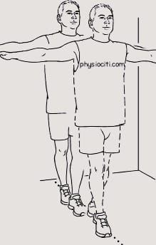
Heel-to-Toe Walking: Walk in a straight line, placing the heel of one foot directly in front of the toes of the opposite foot with each step.
6. Ankle Mobility Exercise
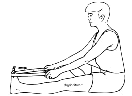
Towel Stretch: Sit with your legs extended. Loop a towel around the ball of your foot and gently pull it towards you, feeling a stretch in your calf and ankle.
7. Functional Exercises
Step-Ups: With your injured leg, step up on a step or platform and then step back down.

Calf raises on an Incline: Using the ball of your foot, stand on an inclined surface (such a book or a wedge) and perform calf raises, focusing on various angles.
8. Co-ordination Exercise
Throwing and catching a ball: This drill tests your equilibrium and hand-eye coordination while standing on the injured leg.

Precautions
Avoid Overexertion: Perform exercises within your pain tolerance and avoid pushing through severe discomfort.
Gradual Progression: As instructed by your physiotherapist or healthcare professional, begin with simpler exercises and work your way up to more difficult ones.
Rest When Needed: To avoid putting too much tension on the recovering ankle, give yourself enough time to recover between workouts.
Before beginning any fitness program following a Pott’s fracture, always check with your doctor or physiotherapist to be sure the exercises are appropriate for your particular condition and recovery stage.
“It is essential to comprehend the complexity and possible complications of Pott’s fracture in order to identify, diagnose, and treat this kind of ankle injury. Restoring ankle function and guaranteeing a full recovery depend heavily on prompt medical care, precise diagnosis, appropriate treatment, and thorough rehabilitation. Remember receiving timely medical attention from qualified healthcare providers is essential to promoting the best possible recovery and avoiding long-term effects from Pott’s fracture.”
ALL THE BEST!